
Helpline

0300 111 1234

Kidney Stones in ADPKD

This information is for people with autosomal dominant polycystic kidney disease (ADPKD), their families and friends. Kidney stones are common in people with ADPKD. Learn here about causes, symptoms, diagnosis, treatment and prevention.

Key Facts

  • Kidney stones are formed when crystals of salts and minerals group together in the urine.
  • They are also called ‘renal calculi’ or ‘nephrolithiasis’.
  • They can range from the size of gravel to a small pebble.
  • Sometimes they travel from a kidney to the ureter (tube to the bladder) or bladder.
  • Kidney stones are common in people with ADPKD but often don’t cause symptoms.
  • Larger stones may cause severe pain, blood in your wee, or a blockage making it hard to wee.
  • Treatment of kidney stones depends on their size and position.
  • Small stones (less than 6—7 mm) can often be passed when you wee without causing problems or much discomfort.
  • You may need a procedure to break up a large stone so that you can pass it, or have surgery to remove it.
  • Steps to reduce your risk of kidney stones include staying hydrated, limiting salt, and eating a normal amount of calcium.

How common are kidney stones in people with ADPKD?

Kidney stones are more common in people with ADPKD than the general population.

By middle age, roughly 1 in every 10 people with ADPKD have had at least one kidney stone that has caused symptoms. A further 2 in every 10 people with ADPKD have kidney stones that don’t cause any symptoms. These can show up on scans, such as computed tomography (CT).

You might be more likely to get kidney stones as you get older and your ADPKD progresses.

Causes of kidney stones

Kidney stones are formed when crystals of substances found in urine, such as calcium and uric acid, group together. People with ADPKD might be more likely to get kidney stones because:

  • Urine flows more slowly through the kidney, meaning that crystals causing kidney stones have more time to form and group.
  • People with ADPKD can have different urine content:
    • higher levels of stone-forming salts and minerals (oxalate and urea)
    • more acidic urine
    • lower levels of substances that help to stop stones forming (citrate and ammonia)

Symptoms of kidney stones

Kidney stones often cause no symptoms and might be spotted on a scan you have for other reasons. Stones can cause symptoms including severe pain, particularly when they move.

You may experience:

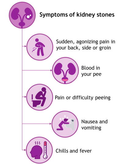
  • sudden, agonizing pain in your back, side or groin (renal colic)
  • blood in your wee
  • pain or difficulty weeing
  • nausea and vomiting
  • chills and fever

You might experience some of these symptoms for other reasons too, such as a cyst infection.

Picture showing the symptoms of kidney stones (as listed in the text above).

Diagnosing kidney stones

Your doctor will examine you and perform some simple tests. These help to work out whether you have a kidney stone or another problem (such as an infected cyst).

These tests usually include:

  • blood tests to check levels of blood cells, your kidney function, and levels of substances that can cause kidney stones
  • urine tests to check for blood in your wee and signs of infection

If your doctor thinks you have a kidney stone, they’ll refer you to a specialist. This should happen within 24 hours if you have severe kidney pain. Alternatively, your doctor may suggest you visit accident and emergency (A + E).

To locate the stone and check its size, you’ll need a scan of your kidneys and bladder, such as a CT scan. A CT machine uses X-rays to develop an image of the inside of your body. You might be given a dye (called contrast medium) before your CT scan to help see any kidney stones. This can be given as a drink or an injection.

Sometimes, other scans may be used, such as:

  • ultrasound, which uses soundwaves to see inside the body
  • intravenous urography (pyelography), for which you have a dye injected into your bloodstream followed by an X-ray

Pregnant women usually have an ultrasound rather than a CT scan or intravenous urography.

Treating kidney stones

Passing small stones in your wee

You might be able to wee out small stones (up to 6—7 mm). Most kidney stones pass in 1—3 weeks at home. During this time, you should increase your fluid intake and may need some treatment including:

  • painkillers (check with your doctor before using non-steroidal anti-inflammatory drug such as ibuprofen or Nurofen®)
  • potassium citrate to try to dissolve the stone or stop it growing
  • drugs to make it easier to pass small stones when you wee (known as alpha blockers)
  • rehydration drinks
  • drugs to stop nausea and vomiting

It’s helpful for doctors to have the kidney stone to test which salts and minerals it’s made of. They can then give you advice or medicine to reduce your chance of having another stone in the future. For this reason, your doctor will ask you to wee through a sieve or gauze to catch the stone and take it to your next clinic visit, if possible.

Procedures to break up larger stones

If your stone is large or you have not been able to wee it out, you may need a procedure to break it up or remove it. This is usually done by a urologist (a doctor specializing in the urinary system) rather than your nephrologist (kidney specialist).

Common procedures are:

  • Shattering the stone with a special machine that delivers shockwaves from outside the body (extracorporeal shockwave lithotripsy). This breaks up the stone into small pieces you can wee out. This technique is commonly used for stones less than 1 cm in size.
  • Using a thin, tube-like camera (a ureteroscope) that is guided through your urethra, bladder and up a ureter (tube to a kidney) to view the stone. A laser is then used to break the stone into smaller pieces (called laser lithotripsy). This is usually done under general anaesthetic. This technique is often used when stones are 1—2 cm in size.
  • ‘Keyhole’ surgery to remove the stone (percutaneous nephrolithotomy). This is done under general anaesthetic through a small cut in the skin.

You should be offered one of these procedures within 48 hours if your pain is ongoing and intolerable, or if the stone is too big to pass naturally.

These procedures generally work well, solving the problem in about 9 out of 10 patients. Lithotripsy sometimes takes more than one session.

The procedures are usually straightforward, but some complications can occur, such as an infection. Ask your doctor to explain the benefits and risks to you, and to outline the different procedures that may be suitable for you. This will help you to make decisions about your treatment.

If you have a large stone and other procedures don’t work, you might need open surgery. This involves a larger cut to access your kidney and is done under general anaesthetic. Very few people need this type of surgery for kidney stones.

Preventing kidney stones

You can reduce your risk of getting kidney stones by:

  • drinking plenty of fluid to avoid dehydration
  • avoiding fizzy drinks
  • adding fresh lemon juice to drinking water
  • eating less salt (no more than 5 g a day)
  • eating a normal amount of calcium (so, having a healthy diet but not using calcium supplements)

      See our diet and lifestyle factsheet for more advice on diet.

      If tests show your kidney stones are caused by a particular salt or mineral, your doctor may recommend that you avoid certain foods. They might also prescribe supplements or medicines to correct imbalances in your wee.

      If you’re taking tolvaptan to slow the progression of your ADPKD, this might also reduce your chance of getting kidney stones. Tolvaptan is not a treatment for kidney stones. However, in a trial of the drug for ADPKD treatment, researchers found that people taking tolvaptan had kidney stones less often than usual. This might be because tolvaptan makes you wee more and reduces the levels of salts and minerals that can cause kidney stones.

      More information from others

      Further Information

      All of our publications are based on references but these are removed for ease of reading on our webpages. A version of this webpage with references included is available upon request by emailing [email protected]

      Authors and contributors

      Written by Hannah Bridges, Independent Medical Writer, HB Health Comms Limited. Expert review by Dr Mattew Gittus, University of Sheffield, UK.

      With thanks to all those affected by ADPKD who contributed to this publication.

      Ref No: ADPKD.KS.V3.0
      Last updated: December 2024
      Next scheduled review: December 2027

      Disclaimer: This information is primarily for people in the UK. We have made every effort to ensure that the information we provide is correct and up to date. However, it is not a substitute for professional medical advice or a medical examination. We do not promote or recommend any treatment. We do not accept liability for any errors or omissions. Medical information, the law and government regulations change rapidly, so always consult your GP, pharmacist or other medical professional if you have any concerns or before starting any new treatment.

      If you don't have access to a printer and would like a printed version of this information sheet, or any other PKD Charity information, call the PKD Charity Helpline on 0300 111 1234 (weekdays, 9am–5pm) or email [email protected]

      The PKD Charity Helpline offers confidential support and information to anyone affected by PKD, including family, friends, carers, newly diagnosed or those who have lived with the condition for many years.

      Feedback

      We welcome feedback on the information provided on this webpage. If you have any feedback regarding the information provided on this webpage, please complete the Feedback Form